690
CEPHALOPODA
membrane, as also is the case in the Dibranchiata. The liver has four paired lobes in Nautilus, which open by two bile-ducts into the alimentary canal at the commencement of the intestine. The bile-ducts unite before entering the intestine. In Dibranchiata the two large lobes of the liver are placed antero-dorsally (beneath the shell in Decapoda), and the bile-ducts open into the caecum. Upon the bile-ducts in Dibranchiata are developed yellowish glandular diverticula, which are known as “pancreas,” though neither physiologically nor morphologically is there any ground for considering either the so-called liver or the so-called pancreas as strictly equivalent to the glands so denominated in the Vertebrata. In Nautilus the equivalents of the pancreatic diverticula of the Dibranchs can be traced upon the relatively shorter bile-ducts.
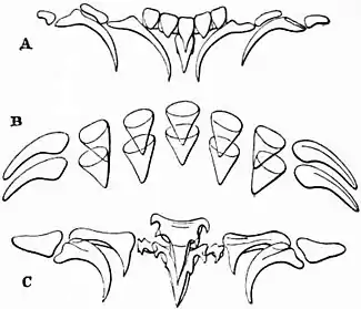 |
| Fig. 9.—Lingual dentition of Cephalopoda. A, A single row of lingual teeth of Nautilus pompilius (after Keferstein). B, Two rows of lingual teeth of Sepia officinalis (after Troschel). C, Lingual teeth of Eledone cirrhosa (after Loven). |
 | |
| Fig. 10.—Diagram representing a vertical approximately median antero-posterior section of Nautilus pompilius (from a drawing by A. G. Bourne). The parts which are quite black are the cut muscular surfaces of the foot and buccal mass. | |
a, The shell.
b, The nuchal plate, identical with the nuchal cartilage of Sepia (see fig. 2, b).
c, The integument covering the visceral hump.
d, The mantle flap or skirt in the dorsal region where it rests against the coil of the shell.
e, The inferior margin of the mantle-skirt resting on the lip of the shell represented by the dotted line.
f, The pallial chamber with two of the four gills.
g, The vertically cut median portion of the mid-foot (siphon).
h, The capito-pedal cartilage (see fig. 8).
i,The valve of the siphon.
l, The siphuncular pedicle (cut short).
m, The hood or dorsal enlargement of the annular lobe of the fore-foot. |
n, Tentacles of the annular lobe.
p, Tentacles of inner inferior lobe.
q, Buccal membrane.
r, Upper jaw or beak.
s, Lower jaw or beak.
t, Lingual ribbon.
x, The viscero-pericardial sac.
n.c, Nerve-collar.
oe, Oesophagus.
cr, Crop.
gizz, Gizzard.
int, Intestine.
an, Anus.
nept, Aperture of a nephridial sac.
r.e, Renal glandular masses on the walls of the afferent branchial veins (see fig. 11).
a.b.v, Afferent branchial vessel.
e.b.v, Efferent branchial vessel.
vt, Ventricle of the heart. |
Posterior salivary glands are not developed in Nautilus, but on each side in the wall of the buccal mass is a gland corresponding to the anterior salivary gland of the Dibranchiata. No ink-sac is present in Nautilus.
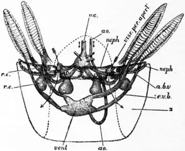 | |
| Fig. 11.—Diagram to show the relations of the four nephridial sacs, the viscero-pericardial sac, and the heart and large vessels in Nautilus (drawn by A. G. Bourne). | |
neph, neph, on the right side point to the two nephridia of that side (the two of the opposite side are not lettered)—each is seen to have an independent aperture.
x is the viscero-pericardial sac, the dotted line indicating its backward extension.
visc.per.apert, marks an arrow introduced into the right aperture of the viscero-pericardial sac. |
r.e, r.e, point to the glandular enlarged walls of the afferent branchial vessels—two small glandular bodies of the kind are seen to project into each nephridial sac, whilst a larger body of the same kind depends from each of the four branchial afferent vessels into the viscero-pericardial sac.
v.c, Vena-cava.
vent, Ventricle of the heart.
ao, Cephalic aorta (the small abdominal aorta not drawn).
a.b.v, Branchial vessel.
e.v.b, Efferent branchial vessel. |