< Page:EB1911 - Volume 06.djvu
This page has been validated.
COCCIDIA
Plate I.
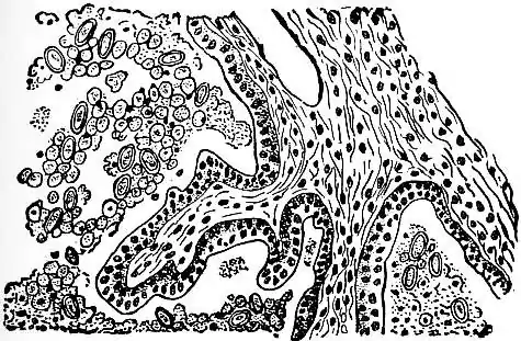 |
| Fig. 1.—SECTION THROUGH RABBIT’S LIVER, INFECTED WITH COCCIDIUM CUNICULI. (AFTER THOMA.) |
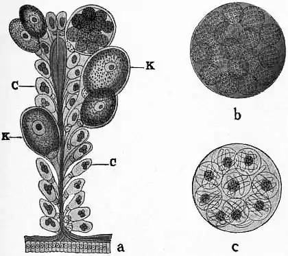 |
| Fig. 2.—KLOSSIA HELICINA, FROM KIDNEY OF HELIX HORTENSIS. |
| a, Portion of a section of the kidney showing normal epithelial cells containing concretions (c), and enlarged epithelial cells containing the parasite (k) in various stages; b, cyst of the Klossia containing sporoblasts; c, cyst with ripe spores, each enclosing four sporozoites and a patch of residual protoplasm. (From Wasielewski, after Balbiani.) |
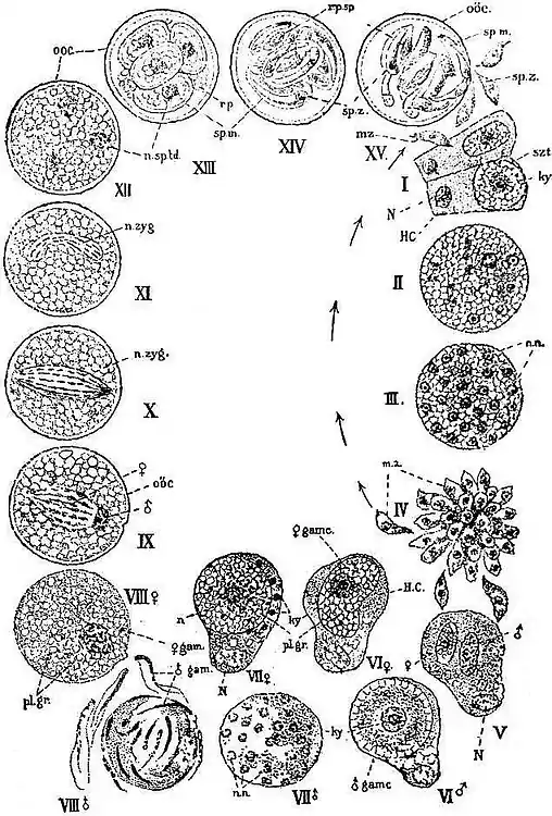 |
| Fig. 3.—THE LIFE-CYCLE OF COCCIDIUM SCHUBERGI, SCHAUD. (PAR. LITHOBIUS FORFICATUS). (FROM MINCHIN, AFTER SCHAUDINN.) |
| I.-IV represents the schizogony, commencing with infection of an epithelial cell by a sporozoite or merozoite. After stage IV the development may start again at stage I, as indicated by the arrows; or it may go on to the formation of gametocytes (V). V-VIII represents the sexual generation. The line of development, hitherto single (I-IV) becomes split into two lines—male (VI ♂, VII ♂, VIII ♂), and female (VI ♀, VII ♀, VIII ♀), culminating in the highly differentiated micro- and mega-gametes. By conjugation these two lines are again united. IX, X, show the formation of the zygote by fusion of the nuclei of the gametes. XI-XV, sporogony. H.C', host-cell; N, its nucleus; mz, merozoite; szt, schizont; ky, karyosome (or fragments of same); n.n, daughter-nuclei of schizont; pl.gr, plastinoid grains; ooc, oocyst; n.zyg, zygote-nucleus (segmentation-nucleus); sp.m, spore-membrane (sporocyst); rp, residual protoplasm of oocyst (“reliquat kystal”); rp.sp, residual protoplasm of spore (“reliquat sporal”); sp.z, sporozoite. |
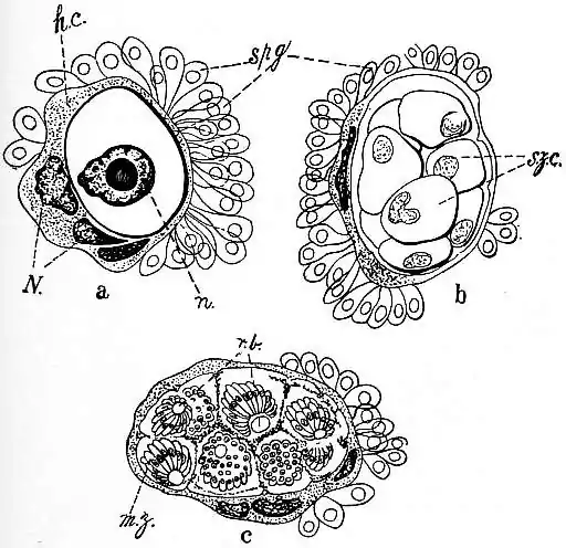 |
| Fig. 4.—PHASES OF CARYOTROPHA MESNILII, SIEDL. (PAR. POLYMNIA NEBULOSA). |
| a, Young schizont in a cluster of spermatogonia; the host-cell (represented granulated) and two of its neighbours are greatly hypertrophied, with very large nuclei, and have fused into a single mass containing the parasite (represented clear, with a thick outline). The other spermatogonia are normal. b, Intracellular schizont divided up into schizontocytes (c), each schizontocyte giving rise to a cluster of merozoites arranged as a “corps en barillet”; spg, spermatogonia; h.c, host-cell; N, nucleus of host-cell or cells; n, nucleus of parasite; szc, schizontocyte; mz, merozoites; r.b, residual bodies of the schizontocytes. (From Minchin, after Siedlecki.) |
This article is issued from Wikisource. The text is licensed under Creative Commons - Attribution - Sharealike. Additional terms may apply for the media files.