LAMELLIBRANCHIA
117
 | |
|
Fig. 12.—Transverse Section of the Outer Gill-plate of | |
| f, | Constituent gill-filaments. |
| ff, | Fibrous sub-epidermic tissue. |
| ch, | Chitonous substance of the filaments. |
| nch, | Cells related to the chitonous substance. |
| lac, | Lacunar tissue. |
| pig, | Pigment-cells. |
| bc, | Blood-corpuscles. |
| fe, | Frontal epithelium. |
| lfe′, lfe″, | Two rows of latero-frontal epithelial cells with long cilia. |
| lrf, | Fibrous, possibly muscular,substance of the inter- filamentar junctions. |
 | |
|
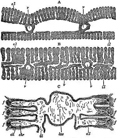
Fig. 13.—Transverse Sections of Gill-plates of Anodonta. | |
| A, | Outer gill-plate. |
| B, | Inner gill-plate. |
| C, | A portion of B more highly magnified. |
| o.l, | Outer lamella. |
| i.l, | Inner lamella. |
| v, | Blood-vessel. |
| f, | Constituent filaments. |
| lac, | Lacunar tissue. |
| ch, | Chitonous substance of the filament. |
| chr, | Chitonous rod embedded in the softer substance ch. |
 |
|
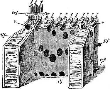
Fig. 14.—Gill-lamellae of Anodonta. (After R. H. Peck.) |
|
Diagram of a block cut from the outer lamella of the outer gill-plate and seen from the interlamellar surface. f, Constituent filaments; trf, fibrous tissue of the transverse inter-filamentar junctions; v, blood-vessel ilj, Inter-lamellar junction. The series of oval holes on the back of the lamella are the water-pores which open between the filaments in irregular rows separated horizontally by the transverse inter-filamentar junctions. |
 | |
|
Fig. 15.—Diagram of a view from the left side of the animal of Anodonta cygnaea, from which the mantle-skirt, the labial tentacles and the gill-filaments have been entirely removed so as to show the relations of the axis of the gill-plumes or ctenidia g, h. (Original.) | |
| a, | Centro-dorsal area. |
| b, | Anterior adductor muscle. |
| c, | Posterior adductor muscle. |
| d, | Mouth. |
| e, | Anus. |
| f, | Foot. |
| g, | Free portion of the axis of left ctenidium. |
| h, | Axis of right ctenidium. |
| k, | Portion of the axis of the left ctenidium which is fused with the base of the foot, the two dotted lines indicating the origins of the two rows of gill-filaments. |
| m, | Line of origin of the anterior labial tentacle. |
| n, | Nephridial aperture. |
| o, | Genital aperture. |
| r, | Line of origin of the posterior labial tentacle. |
shown diagrammatically in fig. 16, C, and more correctly in fig. 17. In this region the inner lamellae of the inner gill-plates are no longer affixed to the foot. Passing still farther back behind the foot, we find in Anodonta the condition shown in the section D, fig. 16. The axes i are now free; the outer lamellae of the outer gill-plates (er) still adhere by concrescence to the mantle-skirt, whilst the inner lamellae of the inner gill-plates meet one another and fuse by concrescence at g. In the lateral view of the animal with reflected mantle-skirt and gill-plates, the line of concrescence of the inner lamellae of the inner gill-plates is readily seen; it is marked aa in fig. 1 (5). In the same figure the free part of the inner lamella of the inner gill-plate resting on the foot is marked z, whilst the attached part—the most anterior—has been snipped with scissors so as to show the genital and nephridial apertures x and y. The concrescence, then, of the free edge of the reflected lamellae of the gill-plates of Anodon is very extensive. It is important, because such a concrescence is by no means universal, and does not occur, for example, in Mytilus or in Arca; further, because when its occurrence is once appreciated, the reduction of the gill-plates of Anodonta to the plume-type of the simplest ctenidium presents no difficulty; and, lastly, it has importance in reference to its physiological significance. The mechanical result of the concrescence of the outer lamellae to the mantle-flap, and of the inner lamellae to one another as shown in section D, fig. 16, is that the sub-pallial space is divided into two spaces by a horizontal septum. The upper space (i) communicates with the outer world