NEUROPATHOLOGY
Plate I.
(Upload an image to replace this placeholder.)
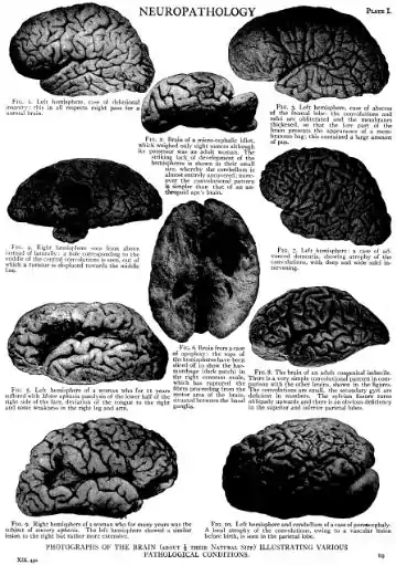
FIG. I. Left hemisphere, case of delusional insanity; this in all respects might pass for normal brain. 4 FIG. 4. Right hemisphere seen from above instead of laterally: a hole corresponding to the middle of the central convolutions is seen, out which a tumour is displaced towards the middle line. FIG. 5. Left hemisiilhere of a woman who for II years suffered with ]l{Dl0f ap sia paralysis of the lower half of the right side of the face, deviation of the tongue to the right and some weakness in the right leg and arm.
FIG. 2. Brain of a micro-cephalic idiot, which weighed only eight ounces although its possessor was an adult woman. The strilting lack of development of the hemispheres is shown in their small size, whereby the cerebellum is almost entirely uncovered; moreover the convolutional pattern is simpler than that of an antllropoid ape's brain. of FIG. 6. Brain fromacase of apoplexy: the tops of the hemispheres have been sliced off to show the hemorrhage (dark patch) in the right centrum ovale, which has ruptured the fibres proceeding from the motor area of the brain, situated between the basal ganglia. PLATE I. FIG. 3. Left hemisphere, case of abscess of the frontal lobe: the convolutions and sulci are obliterated and the membranes thickened, so that the lore part of the brain presents the appearance of a membranous bag; this contained a large amount of pus. FIG. 7. Left hemisphere: a case of advanced dementia, showing atrophy of the convolutions, with deep and wide sulci intervening. FIG. 8. The brain of an adult congenital imbecile. There is a very simple convolutional pattern in comparison with the other brains, shown in the figures. The convolutions are small, the secondary gyri are deficient in numbers. The sylvian fissure turns obliquely upwards and there is an obvious deficiency in the superior and inferior parietal lobes. FIG. Q. Right hemisphere of a woman who for many years was the FIG. IO, Left hemisphere and cerebellum of acase of pore-ncephaly. subjectlof sensory aphasia. The left hemisphere showed a similar A local atrophy of the convolutions, owing to a vascular lesion lesion to the right but rather more extensive. before birth, is seen in the parletal |Ob€-PHOTOGRAPHS OF THE BRAIN (ABOUT § THEIR NATURAL SIZE) ILLUSTRATING VARIOUS PATHOLOGICAL CCNDITIONS. I9
IUX, 4.3o,