PLATE II.
NEUROPATHOLOGY
(Upload an image to replace this placeholder.)
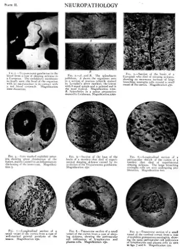
Fig. I.-'I'rypan<>sorna gambicnse in the blood from a case of sleeping sickness in a European. The ilmlulatory membrane is clearly seen; the hcarl of the organism with its micro nucleus is in Contact with a recl blood corpuscle. Magnification 2000 diameters. FIG. 4.-Very marked syphilitic arterit is, showing great diminution of the lumen, mainly caused by an inflammatory thickening of the inner coat. Magnilication 5. FIG. 7.-Longitudinal section of a small vessel of the cortex from a case of well-marked general paralysis of the insane. Magnification 250. B FIG, 2.-/1. and B. The spirochaete pallirlum. A shows the organisms seen in a section of mucous tubercle stained by Leva<liti's silver method; the lowest with 8 equal spirals and a pointed end is the most typical. Magnification moo. B. Spirochetcs in a smear preparation stained by Leishman. Magnification 2260-FIG. 5.-Section of the base of the brain of a monkey that died of experimental sleeping sickness caused by inoculation of the Trypanosoma gambiense. Magnification 250. FIG. 8.-Transverse section of a small vessel of the cortex from a case of sleeping sickness, showing the perivascular cell infiltration of lymphocytes and plasma cells. Magnification 250. FIG. 3.-Section of the brain of a European who died of sleeping sickness, showing an enormous increase of large branching neuroglia cells around a small vessel of the cortex. Magniiication 450. FIG. 6.-Longitudinal section of a perivascular sheath of the cortex of a monkey that died of experimental sleeping sickness. The lar e branching neuroglia cells are seen untiergoing pro life ration. Magnification 600. FIG. 9.-Transverse section of a small vessel of the cerebral cortex from a case of syphilitic gummatous meningitis, show ing the same perivascular cell infiltration of lymphocytes and plasma cells as seen
Ln figs. 7 and 8. Magnification 250.