PARASITIC DISEASES
Plate II.
(Improve this image)
Fig. 5.
Fig. 10.
Fig. 3.
Fig. 7.
Fig. II.
Fig. 4.
Fig. 15. Fig. 16. Fig. 19.
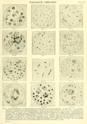
Fig. 2.— -Streptococcus pyogenes, red blood corpuscles and pus cells in the pus from a case of empyaema. (X 1000 diams.) Fig. 3. — Cholera spirillum, from eight days' agar culture, showing many involution forms. Flagella well stained. (X 1000.) Fig. 4. — Bacillus tvphi abdominal is (typhoid bacillus), with well-stained flagella. Young agar cultivation. (X 1000.) Fig. 5. — Group of typhoid bacilli, in a section of spleen. (X 1000.) Fig. 7. — Preparation from young cultivation of Bacillus pestis (plague bacillus). Flagella well stained (X 1000.) Fig. 9. — Bacillus diphtheria, from twenty-four hours' culture. (X 1000.) Fig. 10. — Free edge of false membrane from case of diphtheria containing numerous diphtheria bacilli. (X looo.) Fig. ii. — Bacillus tetani, with well-stained flagella. Twenty-four hours' culture. (X 1000.) Fig. 12. — Scraping from a wound in a case of tetanus, showing several spore-bearing and a few nonspore-bearing tetanus bacilli. (X 1000.) Fig. 15. — Bacillus tuberculosis. Bacilli in a giant-cell in the human liver in a case of acute tuberculosis. (X 1000.) Fig. 16. — Bacillus leprae. Bacilli in endothelial cells of splenic tissue. (X 1000.) Fig. 19. — Amoebae in wall of dysenteric abscess of liver, from specimen kindly lent by Professor Greenfield. (X 1000.)
XX. 774.