152
REPTILES
[ANATOMY
is always formed by the articular bone, not by the angular
which lies on the ventral side, about the middle of the
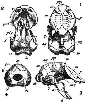 Fig. 22.—Skull of Monopeltis sphenorhynchus.
1, dorsal aspect; 2, ventral aspect; 3, lateral
aspect; 4, posterior aspect. ar, articular; bs,
basisphenoid; d, dentary; f, frontal; m, maxilla;
n, nasal; oc, oc, occipital condyles; of,
occipital foramen; pal, palatine; pa, parietal;
pm, premaxilla; ptg, pterygoid; q, quadrate;
so, supraoccipital; sq, squamosal; v, vomer.
jaw; it is fused
with the articular
in Geckos, some
Tejidae,
Amphisbaenidae, and
some other
burrowing kinds. The
splenial is absent
in chameleons;
near the vanishing
point in some
of the Agamidae.
The coronoid is
always present,
for the insertion
of masseter
muscles. In the
pleurodont lizards
the outer wall of
the dentary forms
a ledge, against
the inner side of
which are fixed
the teeth, with
cementum.
Fig. 22.—Skull of Monopeltis sphenorhynchus.
1, dorsal aspect; 2, ventral aspect; 3, lateral
aspect; 4, posterior aspect. ar, articular; bs,
basisphenoid; d, dentary; f, frontal; m, maxilla;
n, nasal; oc, oc, occipital condyles; of,
occipital foramen; pal, palatine; pa, parietal;
pm, premaxilla; ptg, pterygoid; q, quadrate;
so, supraoccipital; sq, squamosal; v, vomer.
jaw; it is fused
with the articular
in Geckos, some
Tejidae,
Amphisbaenidae, and
some other
burrowing kinds. The
splenial is absent
in chameleons;
near the vanishing
point in some
of the Agamidae.
The coronoid is
always present,
for the insertion
of masseter
muscles. In the
pleurodont lizards
the outer wall of
the dentary forms
a ledge, against
the inner side of
which are fixed
the teeth, with
cementum.
 |
|
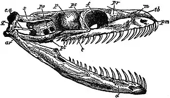
Fig. 23.—Skull of Python sebae. ar, articular; ca, columella auris; d, dentary; f, frontal; m, maxilla; p, parietal; pm, premaxilla; po, proötic; pr, prefrontal; ps, postiiontal; pt, pterygoid; q, quadrate; s, squamosal; t, transversum; tb, turbinal. |
The snakes'
skull shows many
peculiarities, and
most of the bones
of the cranial capsule fuse together without sutures. The
occipital condyle is triple, the lateral occipital and the
basioccipital taking equal share in its composition; the basioccipital
is excluded from the foramen magnum; frequently one common
epiphysial pad covers this tripartite condyle. The supraoccipital
is likewise excluded from the margin of the foramen
magnum by the lateral occipital. The basisphenoid is prolonged
forwards into a long presphenoidal rostrum, on the upper surface
of which the trabeculae cranii, which persist as cartilages,
extend forwards to blend with the median ethmoidal cartilage.
There are no ali- and no orbitosphenoids, their places being
taken by downward extensions of the frontal bones, which
descend to this spheroidal rostrum and then turn inwards
to meet together on the floor of the cranial cavity. There is
consequently no interorbital septum. The parietals also
descend laterally, but unite with the basisphenoid by suture. On
the base of the skull we note various processes for the insertion
of ventral cervicooccipital muscles, much used during the
act of vigorous striking. Boidae have a long spheroidal ridge
and thick basipterygoid processes; others have one or more
median knobs or crests, and the Viperidae have a very
prominent and large ridge. The parietals fuse together into an
unpaired mass whence arises mostly a strong median crest
which projects a little beyond the occiput; there is no parietal
or pineal foramen. There are paired frontals, postfrontals,
 Fig. 24.—Skull of Vipera nasicornis. ar,
articular; ca, columella auris; d, dentary; f,
frontal; m, maxilla; pf, poison fang;
pm, premaxilla; pr, prefrontal; ps, postfrontal;
pt, pterygoid; q, quadrate; s,
squamosal; t, transversum or ectopterygoid.
prefrontals and
nasals; the latter
are said to coössify
in Charina only.
The position of the
prefrontals is
variable. In the boas,
for instance, they
meet, separating the
nasals from the
frontals; they are
in contact with the
nasals in the boas,
burrowing snakes
and in Xenopeltis,
but more or less
widely separated,
from them, and
often from each other, in the Colubridae and Viperidae. The
premaxillary is single; and only in Glauconiidae connected
with the maxillaries; in the others it is but loosely connected
with the ethmoidal end of the skull, for instance, with the
turbinals, which are osseous and well developed in pythons.
Fig. 24.—Skull of Vipera nasicornis. ar,
articular; ca, columella auris; d, dentary; f,
frontal; m, maxilla; pf, poison fang;
pm, premaxilla; pr, prefrontal; ps, postfrontal;
pt, pterygoid; q, quadrate; s,
squamosal; t, transversum or ectopterygoid.
prefrontals and
nasals; the latter
are said to coössify
in Charina only.
The position of the
prefrontals is
variable. In the boas,
for instance, they
meet, separating the
nasals from the
frontals; they are
in contact with the
nasals in the boas,
burrowing snakes
and in Xenopeltis,
but more or less
widely separated,
from them, and
often from each other, in the Colubridae and Viperidae. The
premaxillary is single; and only in Glauconiidae connected
with the maxillaries; in the others it is but loosely connected
with the ethmoidal end of the skull, for instance, with the
turbinals, which are osseous and well developed in pythons.
The whole appendicular apparatus is most loosely attached to the skull, at least in the typical snakes, and since they do not chew their prey but only hook it in, so to speak, during the act of swallowing, the whole apparatus is as movable as possible.
The whole palatal apparatus shows many modifications, but the maxillaries, palatines and pterygoids always remain widely asunder, and from the mid-line. Some of the modifications, so far as they are used for taxonomic purposes, are mentioned in the article Snakes: Classification. In the majority of snakes the maxillaries form the borders of the mouth, and they are but loosely attached to the other bones, to their palatine processes, to the palatines, and with their posterior ends, by the ectopterygoids to the pterygoids. In the Viperidae the maxillaries are much shortened and articulate extensively with the prefrontals; they can be erected, or rather pushed forwards, by the ectopterygoids (see Snakes); they are not connected with the palatines. The pterygoids diverge posteriorly and articulate loosely with the quadrates; in the original condition the articulation is near the distal end of the quadrate, e.g. in Boidae, and the pterygoids may form an additional attachment with the mandibles; in the Viperidae the pterygoids are somewhat shortened and are attached to about the middle of the quadrate shafts; in the Amblycephalidae they are still shorter and do not reach these bones. The ectopterygoids are lost by the burrowing Typhlopidae and Glauconiidae. The quadrate is always extremely movable; besides being in a most curious way connected with the outer end of the columellar rod (see below, Ear), it is suspended from the skull by the squamosal. The squamoso-quadrate connexion is very loose; that of the squamosal with the skull varies much. In the majority of snakes it slides quite freely upon the parietal; it is much longer than the quadrate in the boas, much shorter than the elongated and slender quadrate in most of the poisonous snakes. Lastly, in most of the ancient burrowing snakes, e.g. Typhlops, Glauconia, Ilysia and Uropeltis, the squamosal has worked its way into the cranial wall so that the quadrate, itself also much shortened, rests directly upon the cranium.
The Vertebral Column.
The vertebrae of all reptiles are gastrocentrous, that is to say, the centra or bodies of the vertebrae are formed by the originally paired, interventral cartilages, while the basiventrals are reduced, persisting either as so-called intercentra or wedge-bones, or as intervertebral pads, or disappearing altogether; the basidorsal elements form the neural arch. At the earlier stages of development the gastrocentrous vertebrae behave in the same way as in the Urodela, except that the inter dorsal pair of elements is suppressed from the beginning (the very elements which in